In this manner defects in the afferent or efferent pathways of the light reflex can be established. However, observe the other eye – the other pupil will constrict even without exposure to light (consensual light reflex). First test the direct light reflex – a normal pupil will constrict when light is directed to it.

A normal light reflex results in the constriction of both pupils to light (direct and consensual reflex). Step 2 – Direct and consensual light reflexes. Thus the largest pupil in the light or the smallest pupil in the dark should be the prime suspect in determining which is the abnormal pupil.

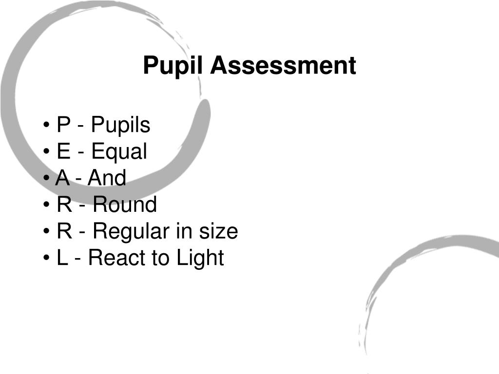
The pathological pupil is the one with the deficient reactivity – either not constricting well to light or dilating poorly in the dark. A greater difference than this is pathological anisocoria. Physiological anisocoria occurs in about 25% of individuals but the difference in size should not be more than 1mm. Get the patient to fix their eyes on a distant point to begin with, then to observe the pupils through a side illumination.Īnisocoria is an inequality in the size of the pupils. A comparison of the size, symmetry and shape of the pupils in both eyes is crucial. Pupils should be examined in light and then in the dark. Step 1 – Compare the sizes of the pupils in the light and the dark. In the far response or in the presence of anxiety, stress or fear, the pupils dilate through this sympathetic activity. This pathway also supplies the Muller’s muscle of the eyelids and the sweat glands of the face. Some of the sympathetic fibres join the ophthalmic division of the trigeminal nerve in the cavernous sinus, then leaves this in the long ciliary nerve to supply the dilator pupillae (Figure 2). Post-ganglionic fibres travel along the external and internal carotid artery. The pre-ganglionic neuron emerges from the first thoracic ventral nerve root to enter the paravetebral sympathetic chain, which runs up to the superior cervical ganglion. The sympathetic pathway starts with the central neuron in the posterior hypothalamus which as it descends is joined in the pons and medulla by the ipsilateral fibres descending from the reticular formation. Pupil dilatation on the other hand is the result of sympathetic activity. The medial recti increase in tone causing the two eyes to converge. At the same time the sphincter pupillae contracts eliminating the passage of light through the peripheral, thinner part of the lens. This results in three responses: the ciliary muscles contract, relaxing the zonules causing the lens to become more globular, increasing the refractive power. Bilateral stimulation from pre-striate cortex area 19 to the Edinger-Westphal nuclei will do the same trick.

Vision is not needed to achieve accommodation. The efferent limb passes from the occipital lobe to the midbrain, where some fibres activate the Edinger-Westphal nucleus as well as the vergence cells in the reticular formation. With accommodation the afferent limb of the reflex passes from the retina to the occipital lobe via the lateral geniculate body. Postganglionic fibres run in the short ciliary nerves and enter the iris to supply the sphincter pupillae (Figure 1). Pre ganglionic parasympathetic fibres enter the oculomotor nerve, leave the branch to the inferior oblique, and synapse in the ciliary ganglion. This pathway results in the direct and indirect light reflex as the input to one optic nerve reaches both Edinger-Westphal nuclei. The contralateral Edinger-Westphal nucleus is reached by way of the posterior commissure. Each pretectal nucleus is linked to its ipsilateral Edinger-Westphal nucleus by internuncial neurons. Fibres leaving the optic chiasm enter both optic tracts and terminate in the pretectal nuclei. The afferent pathway starts in the ganglion cell layer of the retina, which gives rise to the optic nerves. Pupillary constriction is the result of the parasympathetic system activity and is normal in response to two types of stimuli light falling on the retinal photoreceptors and the effort of near reflex and accommodation.Ĭonstriction of the pupils in response to light involves four sets of neurons. The clinical examination of the pupils and pupillary reflexes are crucial in obtaining an accurate diagnosis of a clinical problem.

Pupil size is a result of the interplay between the sympathetic and parasympathetic nervous system supplying the intrinsic muscles within the iris, the dilator and sphincter pupillae respectively. To start at the beginning, the pupil is the central aperture of the iris, its size controlling the amount of light falling on the retina, varying in diameter from about 1-8mm. It is a skill required in eye casualty, clinics and perhaps most importantly, exams. Understanding pupillary reactions is vital in understanding basic neuro-opthalmology.


 0 kommentar(er)
